Hydrocephalus (pronunciation IPA: /ˌhaɪˌdɹoʊˈsɛfələs/) is a condition that can arise in newborns, as well as adults, resulting to swelling of the head which may lead to brain if it can not be treated.
Cerebrospinal fluid (CSF) runs through hollow caves in the nervous system which constitute the ventricular system. In hydrocephalus, it is CSF which causes swelling. CSF normally exist the ventricular system and enters the subarachnoid space where it is absorbed by blood vessels.
CSF is generated in the choroid plexus and it is when the flow from choroid plexus to subarachnoid space is impaired that the ventricles (the canals) begin to swell.
In babies the head expands to accommodate the increase in fluid. If otherwise, the fluid would compress brain tissue leading to brain damage. This is known to occur in adult victims of hydrocephalus because the adult skull can not expand, thus, compression and following brain damage occurs.
To treat hydrophalus a drainage tube is inserted into the lateral ventricle through the skull, allowing for a more regular amount of intracranial fluid.
Anencephaly and Spina bifida
Incompleteness of neural tube formation can result in this fatal condition which occurs very early on in fetal development.
During neurulation, the neural plate is folded and eventually closes. Anencephaly and Spina bifida however, are what can arise when neural tube closure does not complete itself. Depending on which end of the neural tube does not close (anterior or posterior), either condition will arise.
An anterior defect in neurulation results in anencephaly (underdevelopment of the skull and forebrain). A posterior defect will result in Spinal bifeda; a defect of the spine. Whilst anencephaly is always fatal, Spinal bifida can be survived, but only under extensive medical care.
Anencephaly and Spina bifida are believed to be caused by lack of folic acid in diet, which plays a role in DNA biosynthesis which occurs as cells divide.

Anencephaly
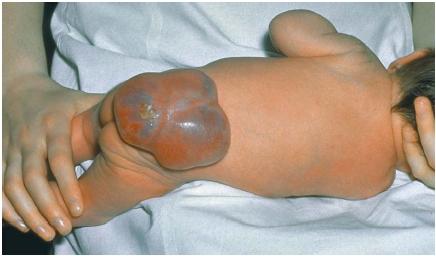
Spina bifida


