I have begun writing my book. It has the core theme of neuroscience and psychology but also includes criminology and sociology. The main objective of the book is to combine knowledge and theories from across fields in the social sciences and to make sense of the world at various levels. For example, how does neuronal functioning lead to prejudice and racism? How does psychology contribute to shaping society? I present answers to these questions by reasoning between fields of study. My answers are not guaranteed 'truths', I present them as possibilities that arise from the implications of findings in particular fields of study.
I am trying to keep the book as simple as possible so that is accessible and interesting to everyone. It serves as an introduction to concepts in the social and biological sciences as well as a tool used to combine them.
If you are interested in how human's shape and are shaped by their worlds; how such tiny and seemingly insignificant matter such as brain cells might give rise to social worlds; or how general features and principles of cognitive psychological processing may shape politics and political decisions, then this book will be of most interest.
Saturday, 30 May 2009
Saturday, 21 March 2009
Neural Correlates of Consciousness
Although there is strong consensus that consciousness and the 'mind' are products of brain activity, there is no evidence of a unifying feature in the brain that 'brings it all together', to produce the 'movie-like' world we experience and this has been on my mind (no pun intended of course) for a while now, while also studying it.
Most studies and experiment to find Neural Correlates of Consciousness (NCCs) are carried out on the visual system. So far, there have been some interesting finds:

All vision does not follow the same path. There are separate streams of vision, cortical (via the cortex) and sub-cortical ('beneath' the cortex). Cortical pathways; the Ventral Stream, are involved in 'higher processing' such as finer details of visual information and the generation of awareness of vision. This is the area of interest here, rather than the Dorsal Stream (sub-cortical pathway) which is inolved in visual processing that occurs without conscious awareness. Yes, we are not consciouslly aware of everything we see, and furthermore, we are not consciouslly aware of everything we sense, but clever experiments can show that unconscious elements of the brain are at work, and pick up things that go unnoticed. Although damage to the primary visual cortex (V1) can cause an indivudal to lose the ability to 'see' objects at a conscious level, the objects do not go unnoticed and this is known as blindsight.
But this does not mean that the primary visual cortex alone constitutes the Ventral stream and gives rise to visual perception. Experiments on monkeys show that the higher degree of neural correlation to conscious perception (the experimenter devised a way to confirm when a monkey was consciouslly perceiving) occurs in the inferotemporal cortex, and that there isn't much correlation in the primary vsual cortex at all. The degree of neural correlation becomes stronger as we move anteriorally along the Ventral stream:
In the inferotemporal cortex we have what are known as essential nodes. These refer to areas of the inferotemporal cortex that are separated by function. In other words, there are different areas for finer detailed visual processing like colour or face recognition. Damage to any one of these specific areas will cause neurological deficeits like prospagnosia (the inability to recognize faces - one famous patient of visual agnosia who showed prospagnosia symptoms is the subject of the book; The Man Who Mistook his Wife for a Hat). Achromatopsia may result from damage to the region that deals with colour.
When these areas are active, we are consciouslly aware of the information they process. It is thus suggested that essential nodes are rather like micro-consciousness'.
The inferotemporal cortex is not the only suspect for NCC in vision.
Damage to the parietal cortex can cause spatial neglect, and this is because spatial awareness is processed in the parietal cortex. There are also prefrontal correlates to visual conscious awareness.
A problem appears to exist. Where as we have subjective experience of unified visual perception, that is, we see the "whole", our visual system is modular. There is no evidence for a space in the brain where everything comes together to generate the unified subjective experience we apparently... experience.
Personally, I don't think we have a unified consciousness. I think consciousness is built of modular, micro-units. Evidence suggest we are aware of colour 80 milliseconds befoe motion. This leads me to believe that the appearance of visual consciousness is just a result of VERY closely related neuronal processes in the visual systems. Breaks occur, but they are so slim we could never notice them. When neurons fire they are not firing continuously- they take very short breaks - so logically, we could never expect a neural correlation of consciousness to show true unification. The location of neurons should be unimportant, just as long as they each serve their own specified and detailed purpose which generates awareness, which I now see as something analogous to disco lights that flicker on and off in short sucession.
Most studies and experiment to find Neural Correlates of Consciousness (NCCs) are carried out on the visual system. So far, there have been some interesting finds:
All vision does not follow the same path. There are separate streams of vision, cortical (via the cortex) and sub-cortical ('beneath' the cortex). Cortical pathways; the Ventral Stream, are involved in 'higher processing' such as finer details of visual information and the generation of awareness of vision. This is the area of interest here, rather than the Dorsal Stream (sub-cortical pathway) which is inolved in visual processing that occurs without conscious awareness. Yes, we are not consciouslly aware of everything we see, and furthermore, we are not consciouslly aware of everything we sense, but clever experiments can show that unconscious elements of the brain are at work, and pick up things that go unnoticed. Although damage to the primary visual cortex (V1) can cause an indivudal to lose the ability to 'see' objects at a conscious level, the objects do not go unnoticed and this is known as blindsight.
But this does not mean that the primary visual cortex alone constitutes the Ventral stream and gives rise to visual perception. Experiments on monkeys show that the higher degree of neural correlation to conscious perception (the experimenter devised a way to confirm when a monkey was consciouslly perceiving) occurs in the inferotemporal cortex, and that there isn't much correlation in the primary vsual cortex at all. The degree of neural correlation becomes stronger as we move anteriorally along the Ventral stream:
In the inferotemporal cortex we have what are known as essential nodes. These refer to areas of the inferotemporal cortex that are separated by function. In other words, there are different areas for finer detailed visual processing like colour or face recognition. Damage to any one of these specific areas will cause neurological deficeits like prospagnosia (the inability to recognize faces - one famous patient of visual agnosia who showed prospagnosia symptoms is the subject of the book; The Man Who Mistook his Wife for a Hat). Achromatopsia may result from damage to the region that deals with colour.
When these areas are active, we are consciouslly aware of the information they process. It is thus suggested that essential nodes are rather like micro-consciousness'.
The inferotemporal cortex is not the only suspect for NCC in vision.
Damage to the parietal cortex can cause spatial neglect, and this is because spatial awareness is processed in the parietal cortex. There are also prefrontal correlates to visual conscious awareness.
A problem appears to exist. Where as we have subjective experience of unified visual perception, that is, we see the "whole", our visual system is modular. There is no evidence for a space in the brain where everything comes together to generate the unified subjective experience we apparently... experience.
Personally, I don't think we have a unified consciousness. I think consciousness is built of modular, micro-units. Evidence suggest we are aware of colour 80 milliseconds befoe motion. This leads me to believe that the appearance of visual consciousness is just a result of VERY closely related neuronal processes in the visual systems. Breaks occur, but they are so slim we could never notice them. When neurons fire they are not firing continuously- they take very short breaks - so logically, we could never expect a neural correlation of consciousness to show true unification. The location of neurons should be unimportant, just as long as they each serve their own specified and detailed purpose which generates awareness, which I now see as something analogous to disco lights that flicker on and off in short sucession.
Tuesday, 10 March 2009
Blood-Brain Barrier
Neuron cells in the central nervous system (CNS)need to be protected against invasion, not only because infection might ensue, but also because their charge may be threatened by ionic compounds that exist in extracelluar space. Thus, cerebral blood vessels are unique in that they are highly restrictive to molecules that circulate in the blood.

The blood-brain barrier is formed by endothelial cells which are cemented together by specialized intercelluar structures called tight junctions. Small molecules such as oxygen (O2) are allowed to pass - and rightly so since of course the brain needs oxygen! What the brain doesn't need is irregular concentrations of ions like potassium or sodium which play a role in keeping cells polarized - readied for action potentials.
The blood-brain barrier is formed by endothelial cells which are cemented together by specialized intercelluar structures called tight junctions. Small molecules such as oxygen (O2) are allowed to pass - and rightly so since of course the brain needs oxygen! What the brain doesn't need is irregular concentrations of ions like potassium or sodium which play a role in keeping cells polarized - readied for action potentials.
Friday, 9 January 2009
Two conditions that can affect brain development...
Hydrocephalus
Hydrocephalus (pronunciation IPA: /ˌhaɪˌdɹoʊˈsɛfələs/) is a condition that can arise in newborns, as well as adults, resulting to swelling of the head which may lead to brain if it can not be treated.
Cerebrospinal fluid (CSF) runs through hollow caves in the nervous system which constitute the ventricular system. In hydrocephalus, it is CSF which causes swelling. CSF normally exist the ventricular system and enters the subarachnoid space where it is absorbed by blood vessels.
CSF is generated in the choroid plexus and it is when the flow from choroid plexus to subarachnoid space is impaired that the ventricles (the canals) begin to swell.
In babies the head expands to accommodate the increase in fluid. If otherwise, the fluid would compress brain tissue leading to brain damage. This is known to occur in adult victims of hydrocephalus because the adult skull can not expand, thus, compression and following brain damage occurs.
To treat hydrophalus a drainage tube is inserted into the lateral ventricle through the skull, allowing for a more regular amount of intracranial fluid.
Anencephaly and Spina bifida
Incompleteness of neural tube formation can result in this fatal condition which occurs very early on in fetal development.
During neurulation, the neural plate is folded and eventually closes. Anencephaly and Spina bifida however, are what can arise when neural tube closure does not complete itself. Depending on which end of the neural tube does not close (anterior or posterior), either condition will arise.
An anterior defect in neurulation results in anencephaly (underdevelopment of the skull and forebrain). A posterior defect will result in Spinal bifeda; a defect of the spine. Whilst anencephaly is always fatal, Spinal bifida can be survived, but only under extensive medical care.
Anencephaly and Spina bifida are believed to be caused by lack of folic acid in diet, which plays a role in DNA biosynthesis which occurs as cells divide.

Anencephaly
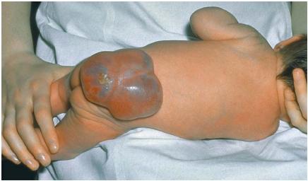
Spina bifida
Hydrocephalus (pronunciation IPA: /ˌhaɪˌdɹoʊˈsɛfələs/) is a condition that can arise in newborns, as well as adults, resulting to swelling of the head which may lead to brain if it can not be treated.
Cerebrospinal fluid (CSF) runs through hollow caves in the nervous system which constitute the ventricular system. In hydrocephalus, it is CSF which causes swelling. CSF normally exist the ventricular system and enters the subarachnoid space where it is absorbed by blood vessels.
CSF is generated in the choroid plexus and it is when the flow from choroid plexus to subarachnoid space is impaired that the ventricles (the canals) begin to swell.
In babies the head expands to accommodate the increase in fluid. If otherwise, the fluid would compress brain tissue leading to brain damage. This is known to occur in adult victims of hydrocephalus because the adult skull can not expand, thus, compression and following brain damage occurs.
To treat hydrophalus a drainage tube is inserted into the lateral ventricle through the skull, allowing for a more regular amount of intracranial fluid.
Anencephaly and Spina bifida
Incompleteness of neural tube formation can result in this fatal condition which occurs very early on in fetal development.
During neurulation, the neural plate is folded and eventually closes. Anencephaly and Spina bifida however, are what can arise when neural tube closure does not complete itself. Depending on which end of the neural tube does not close (anterior or posterior), either condition will arise.
An anterior defect in neurulation results in anencephaly (underdevelopment of the skull and forebrain). A posterior defect will result in Spinal bifeda; a defect of the spine. Whilst anencephaly is always fatal, Spinal bifida can be survived, but only under extensive medical care.
Anencephaly and Spina bifida are believed to be caused by lack of folic acid in diet, which plays a role in DNA biosynthesis which occurs as cells divide.
Anencephaly
Spina bifida
CNS Anatomy Videos
This link will direct you to my youtube channel where I have compiled a playlist of videos instructing CNS anatomy. Anatomy can be difficult to memorize so I recommend using the videos to revise.
Sunday, 4 January 2009
Benzodiazepine and GABA-gated channels
GABA-gated channels mediate inhibition in the Central Nervous System (CNS). Benzodiazephines such as Diazepam bind to the GABA(a) receptor which increases frequency of GABA-gated channel openings, allowing more Cl(-) ions through the channel at synapse, which cause negative charge hyperpolarization, promoting inhibition in the cell. 
Other types of drugs, barbiturates, bind to different sites on the GABA(a) receptor and carry out different tasks which also result in IPSP responses (increasing duration of channel opening). Ethanol also binds to sites on the GABA(a) receptor.
The natural endogenous ligands (binding neurotransmitter) that should resemble benzodiazephine stuctures are yet to be found. They are assumed to exist because receptors for them exist. Similar motivation for the search of endogenous ligands arose from discovery of CB1 and CB2 receptors (cannaboid receptors) where a cannaboid-like ligand was expected to be found (and was found).
Saturday, 3 January 2009
How are neurotransmitters indentified?
The first criterion by which neurotransmitters are recognized as such is that they must be stored and synthesized in the PRE-SYNAPTIC NEURON. With this in mind we turn to two methods of identifying molecules that are stored and synthesized in pre-synaptic neurons, making them worthy neurotransmitter candinates.
Immunocytochemistry is an interesting method with a number of steps. The molecule of interest (the molecule we want to determine whether or not it is neurotransmitter), is injected into the blood of an animal, causing anti-bodies to be released which bind to the 'foreign' substance. In immunology, anti-bodies are specific to chemicals they are deployed against so we can expect anti-bodies that are released to deal with (bind to) an injected substance, let's say, dopamine, to also bind to dopamine if extracted from blood and applied to brain tissue. And this is what actually occurs in the immunocytochemistry technique: the anti-bodies are extracted via withdrawing blood from the animal, and are then tracer-marked (so their activity can be located), and are then applied to brain tissue. The anti-bodies will bind to concentrations of neurotransmitter, giving us localized visual descpritions of the concentration of neurotransmitter. If a neurotransmitter and its synthesizing enzyme is found in the same axon, this helps satisfy the first criterion for idenfication as neurotransmitter.
A second method is In Situ Hybridization, another very interesting method. Here, artifical mRNA is constructed in a labatory with a specific code for binding to mRNA that synthesizes neurotransmitter synthesizing enzymes. The artifical mRNA is labelled with a radioatctive 'tag' and applied to brain tissue. The point in this method is to review locations where neurotransmitter synthesis takes place. Radioactivity of 'probe' mRNA is viewed in autoradiography where locations of synthesis will be revealed. To identify the neurotransmitter, the specific neurotransmitter that the probe mRNA sequence ultimately synthesizes will be reviewed. We have thus identified the neurotransmitter associated with the located synthesizing enzymes.

These methods of identifying neurotransmitter are currently being replaced with more up to date methods of searching for neurotransmitter via receptors.
Immunocytochemistry is an interesting method with a number of steps. The molecule of interest (the molecule we want to determine whether or not it is neurotransmitter), is injected into the blood of an animal, causing anti-bodies to be released which bind to the 'foreign' substance. In immunology, anti-bodies are specific to chemicals they are deployed against so we can expect anti-bodies that are released to deal with (bind to) an injected substance, let's say, dopamine, to also bind to dopamine if extracted from blood and applied to brain tissue. And this is what actually occurs in the immunocytochemistry technique: the anti-bodies are extracted via withdrawing blood from the animal, and are then tracer-marked (so their activity can be located), and are then applied to brain tissue.
The anti-bodies will bind to concentrations of neurotransmitter, giving us localized visual descpritions of the concentration of neurotransmitter. If a neurotransmitter and its synthesizing enzyme is found in the same axon, this helps satisfy the first criterion for idenfication as neurotransmitter.A second method is In Situ Hybridization, another very interesting method. Here, artifical mRNA is constructed in a labatory with a specific code for binding to mRNA that synthesizes neurotransmitter synthesizing enzymes. The artifical mRNA is labelled with a radioatctive 'tag' and applied to brain tissue. The point in this method is to review locations where neurotransmitter synthesis takes place. Radioactivity of 'probe' mRNA is viewed in autoradiography where locations of synthesis will be revealed. To identify the neurotransmitter, the specific neurotransmitter that the probe mRNA sequence ultimately synthesizes will be reviewed. We have thus identified the neurotransmitter associated with the located synthesizing enzymes.
These methods of identifying neurotransmitter are currently being replaced with more up to date methods of searching for neurotransmitter via receptors.
Subscribe to:
Posts (Atom)
